

By Heather Buschman, PhD. For most patients who have died of COVID-19, the pandemic disease caused by a novel coronavirus, the ultimate cause of death was pneumonia, a condition in which inflammation and fluid buildup make it difficult to breathe. Severe pneumonia often requires lengthy hospital stays in intensive care units and assistance breathing with ventilators — medical devices now in high demand in some cities grappling with a surge of COVID-19 cases.
To quickly detect pneumonia — and therefore better distinguish between COVID-19 patients likely to need more supportive care in the hospital and those who could be followed closely at home — UC San Diego Health radiologists and other physicians are now using artificial intelligence (AI) to augment lung imaging analysis in a clinical research study enabled by Amazon Web Services (AWS).
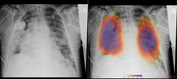
Chest X-rays from a patient with COVID-19 pneumonia, original x-ray (left) and AI-for-pneumonia result (right). Patient has a pacemaker device and an enlarged heart, which indicates that the AI algorithm is powerful enough to work even when the patient has underlying health issues.
The new AI capability has so far provided UC San Diego Health physicians with unique insights into more than 2,000 images. In one case, a patient in the Emergency Department who did not have any symptoms of COVID-19 underwent a chest X-ray for other reasons. Yet the AI readout of the X-ray indicated signs of early pneumonia, which was later confirmed by a radiologist. As a result, the patient was tested for COVID-19 and found to be positive for the illness.
“We would not have had reason to treat that patient as a suspected COVID-19 case or test for it, if it weren’t for the AI,” said Christopher Longhurst, MD, chief information officer and associate chief medical officer for UC San Diego Health. “While still investigational, the system is already affecting clinical management of patients.”
The new capability got its start several months ago when Albert Hsiao, MD, PhD, associate professor of radiology at University of California San Diego School of Medicine and radiologist at UC San Diego Health, and his team developed a machine learning algorithm that allows radiologists to use AI to enhance their own abilities to spot pneumonia on chest X-rays. Trained with 22,000 notations by human radiologists, the algorithm overlays X-rays with color-coded maps that indicate pneumonia probability.
“Pneumonia can be subtle, especially if it’s not your average bacterial pneumonia, and if we could identify those patients early, before you can even detect it with a stethoscope, we might be better positioned to treat those at highest risk for severe disease and death,” Hsiao said.
More recently, Hsiao’s team applied this AI approach to 10 chest X-rays, published in medical journals, from five patients treated in China and the United States for COVID-19. The algorithm consistently localized areas of pneumonia, despite the fact that the images were taken at several different hospitals, and varied considerably in technique, contrast and resolution. The details are published in the Journal of Thoracic Imaging.
Now, enabled by donated service credits provided by the AWS Diagnostic Development Initiative and the efforts of UC San Diego Health’s Clinical Research IT team, Hsiao’s AI method has been deployed across UC San Diego Health in a clinical research study that allows any physician or radiologist to get an initial estimate regarding a patient’s likelihood of having pneumonia within minutes, at point-of-care.
“AWS has partnered with us on multiple projects in the past,” said Michael Hogarth, MD, professor of biomedical informatics at UC San Diego School of Medicine and clinical research information officer at UC San Diego Health. “Once COVID-19 became a crisis, AWS reached out to us and asked if there was anything they could do to help. My mind immediately went to a presentation I’d seen Albert give on their initial AI tests for pneumonia. AWS helped our Clinical Research IT team get the study up and running system-wide in just 10 days.”
According to Hsiao, chest X-rays are cheaper, the equipment is more portable and easier to clean, and results are returned more quickly than many other diagnostics. Polymerase chain reaction-based clinical diagnostic tests for the virus that causes COVID-19 can take several days to return results in some regions of the U.S.
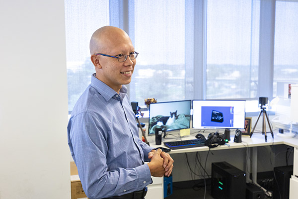
Albert Hsiao, MD, PhD, associate professor of radiology at UC San Diego School of Medicine and radiologist at UC San Diego Health, and team developed a machine learning algorithm that allows radiologists to use AI to enhance their own abilities to spot pneumonia on chest X-rays.
“That’s where imaging can play an important role. We can quickly triage patients to the appropriate level of care, even before a COVID-19 diagnosis is officially confirmed,” Hsiao said.
To be clear, UC San Diego Health experts emphasize they are not diagnosing COVID-19 itself by lung imaging. Pneumonia can be caused by several different types of bacteria and viruses. In addition, use of Hsiao’s AI algorithm is still considered investigational. Although these images are available for use by clinicians, patient care is still guided by formal interpretation from human radiologists.
“As we prepare for a potential surge in patients with COVID-19, it’s not just patient rooms and supplies that may become limited, but also physician and staff capacity,” Longhurst said. “So it’s tremendously helpful to have tools that allow physicians who are not as experienced as radiologists in reading X-rays to get a quick idea of what they’re looking at, especially frontline emergency and hospital-based physicians.”
Next, the UC San Diego Health team hopes to expand the AI-powered study for detecting pneumonia to the University of California’s four other academic medical centers.
“As an academic medical center, we’re always looking for ways to bring innovations to the bedside,” Longhurst said. “Although we need more studies to evaluate the effectiveness of this algorithm and improve its accuracy as we see more patients, what we’re seeing so far is evidence that this approach could be a powerful tool for health care providers to provide more reliable, early diagnoses of COVID-19 and other infections.”
Hsiao’s Journal of Thoracic Imaging study was co-authored by Brian Hurt, MD, and Seth Kligerman, MD, of the Department of Radiology, UC San Diego School of Medicine, and funded in part by the National Institutes of Health (T32 Institutional National Research Service Award), NVIDIA Corporation (GPU grant) and American Roentgen Ray Society.
Disclosure: Albert Hsiao also receives grant support from GE Healthcare and Bayer AG, and is a founder, shareholder, consultant and receives income for Arterys, Inc. Brian Hurt provides consulting services to Arterys, Inc. and IBM.
This article originally appeared in the UC San Diego News Center, April 7, 2020, and is re-posted with permission in the UC IT Blog.